Tag Archives: #metastasis

Breast Cancer’s Response to Tumor Stiffness May Predict Bone Metastasis (Medicine)

In cases of breast cancer, bone metastasis – when cancer cells spread to new sites in the bone – causes the most breast cancer-related harm and is often incurable in advanced disease. A new study by University of Arizona Health Sciences researchers found that cancer cells become more aggressive when exposed to tissue stiffening and that these changes persist over time. 

Tumor stiffening, which develops as diseased breast tissue becomes fibrotic, plays a major role in how breast cancer cells spread throughout the body. The paper, “Breast tumor stiffness instructs bone metastasis via maintenance of mechanical conditioning,” published today in the journal Cell Reports, found that the stiffness of the breast tumor microenvironment can cause changes to cancer cells that make them more aggressively spread to the bone. The resulting changes are maintained as “mechanical memory,” which instructs the cancer cells to send signals that lead to the breakdown of bone. Once this happens, patients often suffer debilitating complications like spontaneous fractures.

Casey Romanoski, PhD, is one of the researchers who helped develop the MeCo score.
Casey Romanoski, PhD, is one of the researchers who helped develop the MeCo score. © UAHS

“Unfortunately, bone metastasis is normally not identified until an advanced state when it’s not reversible,” said senior author Ghassan Mouneimne, PhD, associate professor of cellular and molecular medicine and cancer biology in the UArizona College of Medicine – Tucson. “What’s really exciting is one day being able to take a sample from the patient’s primary tumor and predict who is at high risk for bone metastasis. Then we could intervene with a prevention strategy that we are now validating in the lab.” 

The study, which is the first to demonstrate the concept of mechanical memory during cancer metastasis, developed a novel mechanical conditioning, or “MeCo,” score, to quantify the cellular changes. Eventually, researchers hope the MeCo score can be used to help identify breast cancer patients who might benefit from repurposed antifibrotic treatments to prevent bone metastasis. 

“The higher the patient’s breast tumor MeCo score, the higher the likelihood they would go on to have bone metastasis and poorer outcomes,” said Casey Romanoski, PhD, assistant professor of cellular and molecular medicine and a member of the BIO5 Institute and UArizona Cancer Center. “This stiffness signature could have incredible clinical utility.”

To further explore the clinical application, Dr. Mouneimne and Adam Watson, PhD, a former graduate student and postdoctoral fellow at the UArizona Cancer Center, worked with Tech Launch Arizona, the office of the university that commercializes inventions stemming from research, to launch a startup, MeCo Diagnostics, LLC. The company is working toward maturing the technology and bringing it to the marketplace where it can impact the lives of breast cancer patients everywhere. 

It was previously known that tumor stiffness induces cellular changes that lead to a more aggressive cancer, but according to Dr. Watson, lead author on the paper, the concept of “stiffness” was misleading. 

“Most early-stage breast tumors are stiffer than surrounding tissue, yet most don’t spread to bone,” he said. “It’s not about tumor stiffness but rather stiffness responsiveness of the cancer cells, which we call mechanical conditioning.” 

To study this phenomenon, the team created a laboratory environment that mimicked the stiff or soft tumor environments encountered in the body and assessed how breast cancer cells responded. They found that cells grown in a stiff environment had a “mechanoresponse” characterized by cell spreading, invasion and turning on genes linked with both bone development and disease. And these gene changes endured even after the cells were moved to a soft environment.  

Next, researchers looked at what genes were turned on and off in breast cancer cells in response to the stiff environments. Based on these gene expression changes, they developed the MeCo score, which was validated and refined using data from thousands of patients with breast cancer.

“This is the culmination of a lot of work by researchers from many different fields,” Dr. Mouneimne said. “It highlights the environment we have at the University of Arizona Health Sciences, and how working together can make progress in this challenging area of breast cancer metastasis.”

Future investigations could focus on how cancer cells maintain the gene expression changes that drive metastasis, based on additional findings that identified a transcription factor called RUNX2 that was activated by fibrotic-like stiffness. RUNX2 stays attached to the DNA as the cell divides and “bookmarks” which genes remain turned on, which includes the genes that drive bone metastasis and the breakdown of bone.

Featured image: Ghassan Mouneimne, PhD, hopes the findings of a recently concluded study will lead to a way to predict which breast cancer patients are at high risk for bone metastasis. © UAHS


Provided by University of Arizona Health Sciences
Small Numbers of Cells in a Tumor Could Be Key Enablers of Cancer Metastasis (Medicine)

Just a small number of cells found in tumors can enable and recruit other types of cells nearby, allowing the cancer to spread to other parts of the body, report Georgetown Lombardi Comprehensive Cancer Center scientists. Working with their research collaborators, the scientists found that “enabler cells” comprise about 20 percent or less of the cells in an aggressive tumor; their small numbers may account for why they are often missed when bulk tissue analyses are used to inform therapeutic decisions.

The finding appears online June 16, 2021, in Cancer Research, a journal of the American Association for Cancer Research.

“Our novel finding goes beyond the common understanding of cancer progression as one modeled on Darwinian selection where ‘survival of the fittest’ means the predominant type of cell in a tumor dictates its outcome,” noted Anna Riegel, PhD, professor of oncology and pharmacology at Georgetown Lombardi and the corresponding author of the study. “This could have major implications for our understanding of how best to diagnosis and treat certain cancers, as blocking key cancer-promoting subpopulations of cells might be a way to defeat the cancer.”

The advent of advanced gene sequencing technology, coupled with the use of CRISPR, a tool that allows for easy gene editing, made this finding possible; it is a collaborative effort with researchers at The Ohio State University and Hackensack University Medical Center’s John Theurer Cancer Center, a part of Georgetown Lombardi Comprehensive Cancer Center. These tools aided the scientists in building on their knowledge of alternative splicing, or cutting, of genes whereby a single gene can be spliced to code for multiple proteins.

The researchers’ work using CRISPR in both zebrafish and mice zeroed in on cell subpopulations responsible for enabling metastasis. This led researchers to the discovery of a single RNA splicing event in the AIB1 (amplified in breast cancer 1) gene. One splice variant of the gene produced the AIB1-Delta4 protein, which was found to be responsible for promoting communication and recruitment of surrounding cells, eventually leading to metastasis.

“We propose that the detection of these enabler cells in early-stage breast cancers could predict which tumors are more aggressive and destined to metastasize,” said Ghada M. Sharif, PhD, research assistant professor at Georgetown Lombardi and first author of this finding. “Therapeutic targeting of vulnerabilities uncovered in the enabler cells, such as the splice variants, could represent a new approach to preventing malignant progression of breast cancer.”

This finding is particularly relevant in triple-negative breast cancers which can be aggressive and hard to treat. These types of cancer usually start as non-malignant tumors, called ductal carcinoma in situ (DCIS), but in about 5 to 10 percent of women, they can quickly progress to malignant tumors. The investigators found that AIB1-Delta4 is found at increased levels in women with higher-risk DCIS.

The researchers’ next step will be to conduct various single cell analyses in human tissue samples. “We are at a turning point in how we analyze tumor samples,” said Riegel. “It was unthinkable and impractical just a few years ago to look at every single cell in a tissue sample. But technology is racing ahead, and we believe that in the next few years, looking at each cell will allow us to determine which cells, even if they are small in number, are truly driving cancer progression.”

In addition to Riegel and Sharif, authors of the manuscript at Georgetown include Apsra Nasir, Surojeet Sengupta, Garrett T. Graham, Max H. Kushner, William B. Kietzman, Marcel O. Schmidt, Gray W. Pearson and Anton Wellstein. Other authors include Moray J. Campbell at The Ohio State University, Columbus, OH; Olivier Loudig at the Hackensack Meridian Health Center for Discovery and Innovation, Nutley, NJ; and Susan Fineberg at the Albert Einstein College of Medicine of Yeshiva University, Bronx, NY.

The authors report having no personal financial interests related to the study.

This research was supported by grants from the National Cancer Institute (R01CA205632, R01CA218670, R21CA226542, T32CA009686, F31CA232664).

Featured image: An illustration of a cancer cell migrating through a blood vessel. New research by Georgetown Lombardi scientists and others indicates that just a small number of cells found in tumors can enable and recruit other types of cells nearby, allowing the cancer to spread to other parts of the body. (Image: Annie Cavanagh / Wellcome Collection. Attribution-NonCommercial 4.0 International (CC BY-NC 4.0) )


Reference: Ghada M Sharif, Moray J Campbell, Apsra Nasir, Surojeet Sengupta, Garrett T Graham, Max H Kushner, William B Kietzman, Marcel O Schmidt, Gray W. Pearson, Olivier Loudig, Susan Fineberg, Anton Wellstein and Anna T. Riegel, “An AIB1 isoform alters enhancer access and enables progression of early stage triple-negative breast cancer”, Cancer Research, 2021. DOI: 10.1158/0008-5472.CAN-20-3625


Provided by GLCCC

Study Reveals How Y-box Binding Protein 1 Promotes Cancer Metastasis (Medicine)

Metastasis of cancer is the main cause of cancer relevant death as well as the main challenge of cancer treatment.

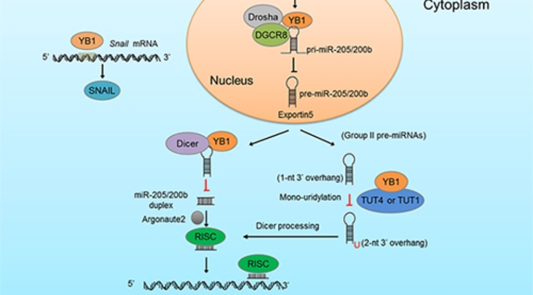
Recently, a group led by Prof. PIAO Hailong from the Dalian Institute of Chemical Physics (DICP) of the Chinese Academy of Sciences (CAS) revealed that the nucleotide binding protein Y-box binding protein 1 (YB1) can promote the metastasis of liver cancer by regulating the biosynthesis of microRNA (miRNA).

This study was published in Cancer Communications on June 10.

The researchers found that YB1 could significantly inhibit the biosynthesis and expression of miRNA-200b and miRNA-205 by interacting with miRNA microprocessor complex DgCR8, Dicer and terminal UTP transferase TUTs. Then, it up-regulated the expression of zinc-finger E-box binding homeobox 1 (ZEB1), which is a key protein for cancer metastasis.

The development of lung cancer metastasis was simulated by a mouse model injected through tail vein, and the researchers found that YB1 could promote lung metastasis of hepatocellular carcinoma cells.

Furthermore, transcriptomics data of clinical tissues showed that the expression of YB1 was positively correlated with ZEB1 in liver cancer tissues, and negatively correlated with the expression of miRNA200 and miRNA205b. The miRNA200/205b-ZEB1 signaling pathway was significantly correlated with the prognosis of patients.

This work was supported by the National Natural Science Foundation of China, Innovation program of science and research from the DICP, and the construction of Liaoning Provincial Cancer Center.

Featured image: YB1 triggers cell invasion and cancermetastasis by regulating the miRNA200/205b-ZEB axis (Image by LIU Xiumei and CHEN Di) 


Reference: Liu, X, Chen, Di, Chen, H, Wang, W, Liu, Yu, Wang, Y, et al. YB1 regulates miR-205/200b-ZEB1 axis by inhibiting microRNA maturation in hepatocellular carcinoma. Cancer Commun. 2021; 1– 20. https://doi.org/10.1002/cac2.12164


Provided by Chinese Academy of Sciences

Mutant KRAS and p53 Cooperate To Drive Pancreatic Cancer Metastasis (Medicine)

Preclinical research identifies CREB1 as new therapeutic target downstream of frequently mutated genes

Researchers at The University of Texas MD Anderson Cancer Center have discovered that mutant KRAS and p53, the most frequently mutated genes in pancreatic cancer, interact through the CREB1 protein to promote metastasis and tumor growth. Blocking CREB1 in preclinical models reversed these effects and reduced metastases, suggesting an important new therapeutic target for the deadly cancer.

The findings were published today in Cancer Discoveryand presented at the virtual American Association for Cancer Research (AACR) Annual Meeting 2021 by Michael Kim, M.D., assistant professor of Surgical Oncology and Genetics.

“To our knowledge, this is the first study to show how these two major genetic drivers work together to promote tumor growth and metastasis,” Kim said. “We learned that signaling downstream of mutant KRAS directly promotes mutant p53 activity. This discovery provides not only a new therapeutic target but unveils a vast transcriptional network that is activated downstream of these mutant proteins.”

Mutations in KRAS and TP53, the two most frequently mutated genes in all human cancers, co-occur in roughly 70% of patients with pancreatic cancer. Mutant KRAS, found in 95% of pancreatic cancers, leads to an activated protein that aberrantly triggers many downstream signaling pathways. Mutant TP53 results in the loss of the proteins’ tumor suppressor function, leaving the mutant protein capable of fueling additional oncogenic processes, such as metastasis.

Unfortunately, no current therapies are able to block the mutant forms of KRAS or p53 prevalent in pancreatic cancer, so there is a need to identify common, alternative therapeutic targets downstream of these proteins that could lead to more effective treatment regimens for pancreatic cancer, Kim explained.

To learn how mutant KRAS and p53 might be interacting, Kim’s team of researchers collaborated with Gigi Lozano, Ph.D., chair of Genetics, to develop a novel mouse model of pancreatic cancer that expresses oncogenic KRAS and mutant p53 specifically in tumor cells, leaving the tumor microenvironment unaltered.

In this model, the team observed more than twice as many metastatic lesions than was seen when p53 was genetically removed, suggesting that the mutant proteins together cause a significant increase in metastatic potential. With further study, the researchers discovered mutant KRAS activates CREB1, a transcription factor that then directly interacts with mutant p53 to promote the aberrant expression of hundreds of genes.

Michael Kim, M.D. © MD Anderson Cancer Center

This activation results in the increased expression of FOXA1, which in turns creates a new cascade of events leading to increased activity of the Wnt/β-catenin pathway, both of which promote cancer metastasis.

Using an available small-molecule drug to target CREB1 in this model resulted in decreased expression of FOXA1β-catenin and associated target genes, along with a corresponding reduction in metastases. While early, these findings suggest that targeting CREB1 may be a viable strategy to block the metastatic effects of mutant KRAS and p53 in pancreatic cancer.

“The identification of this cooperative node suggests that there should be increased focus on CREB1 as a target that could be therapeutically exploited to improve patient outcomes,” Kim said. “With the frequency of KRAS and TP53 mutations in human cancers, the implications of our findings may extend far beyond pancreatic cancer.”

Going forward, the researchers hope to discover other important elements working downstream of mutant p53 that may affect the cancer cells or the surrounding tumor microenvironment. A greater understanding of this complex network may point to additional therapeutic targets or combination approaches to better treat pancreatic cancer.

The research was supported by the National Institutes of Health (NIH) (K08CA218690, P01CA117969, R01CA82577, T32 CA 009599, 1S10OD024976-01, P30CA16672), the American College of Surgeons Faculty Research Fellowship, the Cancer Prevention & Research Institute of Texas (CPRIT) (RP17002), the Richard K. Lavine Pancreatic Fund, the Ben and Rose Cole Charitable Pria Foundation, and the Skip Viragh Foundation.

In addition to Kim, MD Anderson collaborators on the study include: Xinqun Li, M.D., Ph.D., Jenying Deng, Ph.D., Bingbing Dai, Ph.D., Tara G. Hughes, M.D., Christian Siangco, Jithesh Augustine, and Yaan Kang, M.D., all of Surgical Oncology; Yun Zhang, Ph.D., Joy M. McDaniel, Ph.D., Shunbin Xiong, Ph.D., Amanda R. Wasylishen, Ph.D., and Guillermina Lozano, Ph,.D., all of Genetics; Kendra Allton, Bin Liu, Ph.D., and Michelle C. Barton, Ph.D., all of Epigenetics and Molecular Carcinogenesis; Eugene Koay, M.D., Ph.D., of Radiation Oncology; Florencia McAllister, M.D., of Gastrointestinal Medical Oncology and Clinical Cancer Prevention; Christopher Bristow, Ph.D., and Timothy P. Heffernan, Ph.D., of the TRACTION platform; and Anirban Maitra, M.B.B.S., of the Sheikh Ahmed Bin Zayed Al Nahyan Center for Pancreatic Cancer Research. Additional co-authors include Jason B. Fleming, M.D., of Moffitt Cancer Center and Research Institute, Tampa, FL. The authors declare no conflicts of interest.

Featured image credit: Image courtesy Michael Kim, M.D.


References: (1) Michael P Kim, Xinqun Li, Jenying Deng, Yun Zhang, Bingbing Dai, Kendra L Allton, Tara G. Hughes, Christian Siangco, Jithesh J. Augustine, Ya’an Kang, Joy M McDaniel, Shunbin Xiong, Eugene J Koay, Florencia McAllister, Christopher A. Bristow, Timothy P. Heffernan, Anirban Maitra, Bin Liu, Michelle C. Barton, Amanda R Wasylishen, Jason B. Fleming and Guillermina Lozano, “Oncogenic KRAS recruits an expansive transcriptional network through mutant p53 to drive pancreatic cancer metastasis”, Cancer Discovery, 2021. DOI: 10.1158/2159-8290.CD-20-1228 (2) Michael Paul Kim, Xinqun Li, Jenying Deng, Yun Zhang, Bingbing Dai, Kendra Allton, Tara Hughes, Christian Siangco, Jithesh Augustine, Yaan Kang, Joy M. McDaniel, Shunbin Xiong, Eugene Koay, Florencia McAllister, Christopher Bristow, Timothy Heffernan, Anirban Maitra, Bin Liu, Michelle Barton, Amanda Wasylischen, Jason Fleming, Guillermina Lozano, “Mutant p53 and oncogenic KRAS converge on CREB1 to drive pancreatic cancer metastasis”, Session PO.MCB03.05 – Nuclear Oncoproteins, Oncogenes, and Tumor Suppressor Genes, 2021. ABSTRACT #2417


Provided by MD Anderson Cancer Center

Creatine Promotes Cancer Metastasis Through Activation of Smad2/3 (Medicine)

As one of the most popular nutrient supplements, creatine has been used to enhance muscle mass and the function of heathy human subjects. Dietary creatine supplementation has even been used in clinical trial to reverse cachexia of colorectal cancer (CRC) patients, although creatine uptake has been demonstrated to fail to improve either muscle function or life quality of the patients.Studies using subcutaneous injection mouse models showed that creatine uptake suppresses tumor growth, setting creatine as an anti-tumor supplement. However, subcutaneous injection mouse models are not clinically relevant for investigation of tumor progression, and tumor growth is not always coupled with metastasis. Thus, it remains to be elucidated how creatine affects tumor progression, such as metastasis and patient survival.

In a study published in Cell Metabolism, a research group led by Prof. BU Pengcheng at the Institute of Biophysics of the Chinese Academy of Sciences (CAS), collaborating with PIAO Hailong from Dalian Institute of Chemical Physics of the CAS and CHEN Gang from Chinese PLA General Hospital, reported that creatine promotes cancer metastasis through activation of Smad2/3.

To explore creatine function in cancer metastasis, the researchers used an orthotopic mouse model and found that dietary uptake creatine promoted the metastasis of colorectal cancer and breast cancer and shortened mice survival.

They found that the rate-limiting enzyme for creatine synthesis, glycine acyltransferase (GATM), is up-regulated in liver metastasis. Clinically, high levels of GATM were significantly associated with poorer prognosis. GATM promoted CRC liver metastasis and shortened mouse survival, while targeting GATM or creatine transporter SLC6A8 reduced CRC liver metastasis and prolonged mouse survival.

To investigate the mechanism, the researchers performed multi-pathway profiling array and found that creatine upregulated Smad2/3 phosphorylation. Creatine activated Smad2/3 signaling via monopolar spindle 1 (MPS1), a protein kinase associated with mitotic progression and spindle checkpoint.

They further found that creatine also upregulated downstream transcription factor Snail/Slug expression. MPS1 knockdown and specific inhibitor inhibited primary tumor invasion and cancer metastasis by down-regulating Snail/Slug expression, and prolonged mice survival.

This study called for using caution when considering dietary creatine to improve muscle mass or clinical treatment especially for cancer patients. It reported creatine activated Smad2/3 through MPS1, rather than transforming growth factor beta receptor (TGFBR) in the canonical TGF-beta pathway. They also suggested that targeting GATM or MPS1 prevents cancer metastasis, especially metastasis of TGFBR mutant colorectal cancers.

CRC is the third most deadly and fourth most commonly diagnosed cancer in the world, and the incidence has been steadily rising. About 25% of diagnoses cases has metastasized and 70% of CRC patients eventually develop liver metastasis. Hereditary CRC account for 7-10% of all cases, and diet are significant CRC risk factors.


Reference: Liwen Zhang, Zijing Zhu, Huiwen Yan, Wen Wang, Zhenzhen Wu, Fei Zhang, Qixiang Zhang, Guizhi Shi, Junfeng Du, Huiyun Cai, Xuanxuan Zhang, David Hsu, Pu Gao, Hai-long Piao, Gang Chen, Pengcheng Bu, Creatine promotes cancer metastasis through activation of Smad2/3, Cell Metabolism, 2021, , ISSN 1550-4131, https://doi.org/10.1016/j.cmet.2021.03.009. (https://www.sciencedirect.com/science/article/pii/S1550413121001169)


Provided by Chinese Academy of Sciences

CRISPR study identifies gene that plays key role in metastasis of cancers to the lungs

Over-expression of LRRN4CL was linked to cancer spread in colon, breast and bladder cancers in humans

A gene not previously linked to cancer has been shown to play a key role in the spread of certain cancers to the lungs, new research from scientists at the Wellcome Sanger Institute has shown. The team found that when the gene LRRN4CL was over-expressed in mice, the skin cancer melanoma was more likely to metastasise to the lungs.

The study, published today (23 March 2021) in Communications Biology, also confirmed that over-expression of LRRN4CL was linked to metastasis of colon, breast and bladder cancers to the lung.

Several factors make LRRN4CL an attractive drug target. It encodes a protein found on the surface of cancer cells, making it easier to target with drugs. And because it is expressed at low levels elsewhere in the body, it may be possible to target LRRN4CL without causing serious side effects for the patient.

Metastasis is when cancers spread from one organ to other parts of the body via the blood and lymphatic systems. Once cancer cells are mobile, they can take root in other organs and form secondary tumours, most commonly in the lungs.

Metastasis makes cancers much harder to treat and is associated with much poorer outcomes for patients. Metastasis is the reason why melanoma, which accounts for around 5 per cent of skin cancers, also accounts for the majority of skin cancer deaths. Whereas basal cell skin cancer, which is the most common form of the disease, rarely spreads and is rarely life-threatening*.

In this study, scientists at the Wellcome Sanger Institute set out to identify genes whose over-expression – an increase in activity compared to the gene’s normal functioning – resulted in the increased ability of melanoma cells to spread to the lungs in mice.

They conducted a CRISPR activation (CRISPRa) screen on genes that encode proteins on the surface of the cell, boosting the level of expression of the genes one by one to observe which had an effect.

The team found that over-expression of the gene LRRN4CL increased the ability of mouse and human melanoma cells to metastasise to the lungs. They also tested cell models for colon, breast and bladder cancer, confirming the role of LRRN4CL in metastasis to the lungs in all of them. This is the first time that LRRN4CL has been linked to cancer.

“Metastasis is a complex phenomenon, and metastatic cells can have different characteristics depending on the original cancer type, the secondary tumour location and even the age of the patient. However, our findings show that when metastatic cancer cells of the skin, colon or breast express high levels of LRRN4CL, it makes them uniquely able to survive and grow in the lung.”

— Dr Louise van der Weyden, first author of the paper from the Wellcome Sanger Institute

The researchers also checked databases of gene expression in patient tumours and found that melanomas with elevated expression of LRRN4CL correlated with poorer patient outcomes.

“Our results suggest that reducing the expression of the LRRN4CL gene could help to prevent metastasis to the lungs, which would already make it a potential drug target. The added bonus is that this gene is expressed at very low levels elsewhere in the body, so hopefully targeting LRRN4CL wouldn’t have severe side effects for patients.”

Dr Anneliese Speak, an author of the paper from the Wellcome Sanger Institute

Genes that encode cell surface proteins are often attractive drug targets, as the drug only needs to be delivered to the exterior of the cell rather than the interior, making drug design less complex.

“Prior to this study, there was nothing in the scientific literature to link the LRRN4CL gene to cancer, much less to suggest that it plays such a pivotal role in metastasis. Part of the power of CRISPR screens is that they don’t require a clear hypothesis to create new insights. This is an important discovery that marks LRRN4CL as a promising drug target to help prevent the spread of cancer to the lungs and improve outcomes for patients.”

Dr Dave Adams, senior author of the paper from the Wellcome Sanger Institute

In additional work recently published in Nature Communications, the team used combinatorial CRISPR screening to define gene pairs that represent therapeutic targets in melanoma, thus providing additional targets to treat this complex disease.

Publication:

Louise van der Weyden, Victoria Harle and Gemma Turner et al. (2021). CRISPR activation screen in mice identifies novel membrane proteins enhancing pulmonary metastatic colonisationCommunications Biology. DOI: 10.1038/s42003-021-01912-w

https://doi.org/10.1038/s42003-021-01912-w

Funding:

This work was funded by Cancer Research UK, ERC Combat Cancer and Wellcome.

Featured image credit: Anne Weston, Francis Crick Institute


Provided by Wellcome Sanger Institute

Researchers Reveal Mechanism of Hepatitis B-induced Venous Metastasis & Immune Escape of Hepatocellular Carcinoma (Medicine)

In China, about 70 million people are infected with hepatitis B virus (HBV), and more than 80% of liver cancer is caused by HBV.

HBV-associated hepatocellular carcinoma (HCC) is often accompanied by severe vascular invasion and portal vein tumor thrombus leading to a poor prognosis. However, the underlying mechanism of this disease remains obscure.

In a study published in Cancer Research, Prof. YANG Pengyuan and Prof. WANG Fan at the Institute of Biophysics (IBP) of the Chinese Academy of Sciences (CAS) revealed how HBV infection induced HCC venous metastasis and immune escape through a chemokine-based network.

The researchers firstly established a cytokines/chemokines array and screened the proinflammatory cytokine IL-8 as a potential target responsively to HBV infection. Multiple models and mechanistic study identified that HBV-induced IL-8 expression can be activated by HBx-mediated MEK-ERK signaling pathway, which enhanced permeability of the endothelium via endothelial CXCR1, the receptor of IL-8.

Based on the identification of IL-8-CXCR1 axis promoting tumor vascular metastasis in vivo, they then constructed a transgenic mouse that selectively express human CXCR1 in endothelial cells.

Interestingly, the IL-8-CXCR1 axis on vascular endothelium dramatically increased liver tumorigenesis and metastasis, and the increase in lung metastasis was observed through overexpression of IL-8, but exogenous CXCR1 overexpression did not further enhance lung metastasis. Mechanistically, the IL-8-CXCR1 axis selectively induced GARP-latent-TGF-β in liver sinusoidal endothelial cells (LSECs) and subsequently provoked preferential regulatory T cell polarization to suppress antitumor immunity.

This study identifies a hepatitis B-induced IL-8/CXCR1/TGF-β signaling cascade that suppresses anti-tumor immunity and enhances metastasis in hepatocellular carcinoma, providing new potential targets for therapeutic intervention.

Featured image: Working model of IL-8-CXCR1 axis promoted venous metastasis and intrahepatic Treg accumulation in HBV-associated HCC (Image by Dr. YANG Pengyuan’s group)


Reference: Changlu Zhang, Yanan Gao, Chengzhi Du, Geoffrey J. Markowitz, Jing Fu, Zhenxing Zhang, Chunliang Liu, Wenhao Qin, Hongyang Wang, Fan Wang and Pengyuan Yang, “Hepatitis B-induced IL-8 Promotes Hepatocellular Carcinoma Venous Metastasis and Intrahepatic Treg Accumulation”, Cancer Res March 2 2021 DOI: 10.1158/0008-5472.CAN-20-3453


Provided by Chinese Academy of Sciences

New Insight Into How Cancer Spreads (Medicine)

Heide Ford, PhD, associate director of basic research at the CU Cancer Center, had a study published in Oncogene investigating how cancer metastasizes.

Breast cancer is harmful enough on its own, but when cancer cells start to metastasize — or spread into the body from their original location — the disease becomes even more fatal and difficult to treat.

Thanks to new research published in Oncogene from the lab of University of Colorado Cancer Center associate director of basic research Heide Ford, PhD, in collaboration with Michael Lewis, PhD, from Baylor College of Medicine, doctors may soon have a better understanding of one mechanism by which metastasis happens, and of potential ways to slow it down.

“Metastasis is a huge problem nobody’s tackled very well,” says Ford, who holds the Grohne Endowed Chair in Cancer Research at the University of Colorado School of Medicine. “People don’t know how to inhibit the process of metastasis, nor how to inhibit the growth of metastatic cells at secondary sites. And that’s what kills most cancer patients. A lot of common drugs, whether they’re targeted drugs or chemotherapies that are less targeted, do pretty well at inhibiting the primary tumor, but by the time cells metastasize, they’ve changed enough that they don’t get inhibited by those drugs.”

The epithelial-to-mesenchymal transition

The transformation Ford and her team are studying happens when cells called epithelial cells, which are more adherent to one another and less likely to spread to other parts of the body, start to take on the characteristics of mesenchymal cells, which are more migratory and more likely to invade other parts of the body. This transformation is referred to as the epithelial-to-mesenchymal transition.

Heide Ford, PhD

“When the epithelial cancer cells take on these characteristics of mesenchymal cells, they become less attached to their neighbor and they become more able to degrade membranes, so they can get into the bloodstream more easily,” Ford says.

In 2017, Ford published a paper showing that the metastasis process is helped along when cells that have undergone the epithelial-to-mesenchymal transition start “talking” to cells that haven’t, making those cells more likely to gain metastatic properties.

In a new paper published in December, Ford and her researchers, in a collaborative study done with Lewis and colleagues at Baylor College of Medicine, posit that the crosstalk is facilitated by a naturally occurring protein called VEGF-C.

“VEGF-C is secreted by the cells. It binds to receptors on these neighboring cells and then activates a pathway called the hedgehog signaling pathway, though it bypasses the traditional way of activating this pathway,” Ford says. “That turns on a signaling mechanism that ultimately results in activation of a protein called GLI that makes these cells more invasive and more migratory.”

Targeting crosstalk

In their new paper, Ford, Lewis and their researchers show that if you can inhibit production of VEGF-C, you can significantly slow metastasis.

“If you take out the receptor that receives the signal from the cells that have not undergone a transition, or if you take VEGF-C out of the mix, you can’t stimulate metastasis to the same degree,” she says. “If you remove that ability for these different cell types to crosstalk, now these cells that never underwent a transition can’t move as well anymore. They can’t metastasize as efficiently.”

The researchers are now in the early stages of animal trials to find out the best way to target that signaling pathway in order to better inhibit metastasis. They want to find out if they can stop metastasis from happening at all, and if they can slow its progression in patients in whom the metastatic process has already begun — and to see if they can inhibit tumor growth at the secondary site.

“For many years, people said there was no point in finding inhibitors to metastasis because by the time someone comes into the clinic, the horse is out of the barn, so to speak. The cells have already gotten out of the primary tumor and you can’t do anything about it,” Ford says. “But that’s not necessarily true. Now, data show that if you have cells that have metastasized to a second site — say you have breast cancer and the cells went into the lungs — those cells that are in the lungs could in fact start metastasizing to other sites. You want to stop that process no matter where you are in this progression.”


Reference: Kong, D., Zhou, H., Neelakantan, D. et al. VEGF-C mediates tumor growth and metastasis through promoting EMT-epithelial breast cancer cell crosstalk. Oncogene 40, 964–979 (2021). https://www.nature.com/articles/s41388-020-01539-x https://doi.org/10.1038/s41388-020-01539-x


Provided by University of Colorado Cancer Center

Princeton Team Discovers New Organelle Involved in Cancer Metastasis (Biology)

Some of Princeton’s leading cancer researchers were startled to discover that what they thought was a straightforward investigation into how cancer spreads through the body — metastasis — turned up evidence of liquid-liquid phase separations: the new field of biology research that investigates how liquid blobs of living materials merge into each other, similar to the movements seen in a lava lamp or in liquid mercury.

“We believe this is the first time that phase separation has been implicated in cancer metastasis,” said Yibin Kang, the Warner-Lambert/Parke-Davis Professor of Molecular Biology. He is the senior author on a new paper featured on the cover of the current issue of Nature Cell Biology.

Not only does their work tie phase separations to cancer research, but the merging blobs turned out to create more than the sum of their parts, self-assembling into a previously unknown organelle (essentially an organ of the cell).

Discovering a new organelle is revolutionary, Kang said. He compared it to finding a new planet within our solar system. “Some organelles we have known for 100 years or more, and then all of a sudden, we found a new one!”

This will shift some fundamental perceptions of what a cell is and does, said Mark Esposito, a 2017 Ph.D. alumnus and current postdoc in Kang’s lab who is the first author on the new paper. “Everybody goes to school, and they learn ‘The mitochondria is the powerhouse of the cell,’ and a few other things about a few organelles, but now, our classic definition of what’s inside a cell, of how a cell organizes itself and controls its behavior, is starting to shift,” he said. “Our research marks a very concrete step forward in that.”

The work grew out of collaborations between researchers in the labs of three Princeton professors: Kang; Ileana Cristea, a professor of molecular biology and leading expert in the mass spectroscopy of living tissue; and Cliff Brangwynne, the June K. Wu ’92 Professor of Chemical and Biological Engineering and director of the Princeton Bioengineering Initiative, who pioneered the study of phase separation in biological processes.

“Ileana is a biochemist, Cliff is a biophysicist and engineer, and I am a cancer biologist — a cell biologist,” Kang said. “Princeton is just a wonderful place for people to connect and collaborate. We have a very small campus. All the science departments are right next to each other. Ileana’s lab is actually on the same floor of Lewis Thomas as mine! These very close relationships, among very diverse research areas, allow us to bring in technologies from many different angles, and allow breakthroughs to understanding the mechanisms of metabolism in cancer — its progression, metastasis and the immune response — and also come up with new ways to target it.”

The latest breakthrough, featuring the as-yet unnamed organelle, adds new understanding to the role of the Wnt signaling pathway, a system whose discovery led to the 1995 Nobel Prize for Eric Wieschaus, Princeton’s Squibb Professor in Molecular Biology and a professor in the Lewis-Sigler Institute for Integrative Genomics. The Wnt pathway is vital to embryonic development in countless organisms, from tiny invertebrate insects to humans. Wieschaus discovered that cancer can co-opt this pathway, essentially corrupting its ability to grow as rapidly as embryos must, to grow tumors.

This 3D image of human breast cancer bone metastases shows the formation of the newly described organelle (magenta) in cancer cells (cyan). Cell nuclei from both cancer cells and normal bone cells are labeled in blue.
Image rendering by Mark Esposito and Gary Laevsky

Subsequent research has revealed that the Wnt signaling pathway plays multiple roles in healthy bone growth as well as in cancer metastasizing to bones. Kang and his colleagues were investigating the complex interplay between Wnt, a signaling molecule called TGF-b, and a relatively unknown gene named DACT1 when they discovered this new organelle.

Through a series of detailed and complex experiments, the researchers pieced together the story: bone tumors initially induce Wnt signaling, to disseminate through the bone. Then, TGF-b, which is abundant in bones, prompts DACT1 to sequester materials in the new organelle in a way that suppresses Wnt signaling. The tumors then stimulate the growth of osteoclasts, which scrub away old bone tissue. (Healthy bones are constantly being replenished in a two-part process: osteoclasts scrub away a layer of bone, then osteoblasts rebuild the bone with new material.) This further increases the TGF-b concentration, prompting even more DACT1 sequestration and subsequent Wnt suppression that has been shown to be important in further metastasis.

By discovering the roles of DACT1 and this organelle, Kang and his team have found new possible targets for cancer drugs. “For example, if we have a way to disrupt the DACT1 complex, perhaps the tumor will disseminate, but it will never be able to ‘grow up’ to be life-threatening metastasis. That’s the hope,” Kang said.

Kang and Esposito recently co-founded KayoThera to pursue the development of medications for patients with late-stage or metastatic cancers, based on their work together in the Kang lab. “The kind of fundamental study that Mark is doing both presents groundbreaking science findings and can also lead to medical breakthroughs,” said Kang.

The researchers have found that DACT1 plays many other roles as well, which their team is only beginning to explore. The mass spectrometry collaboration with Cristea’s team revealed more than 600 different proteins in the mysterious organelle. Mass spectrometry allows scientists to find out the exact components of almost any substance imaged on a microscope slide.

“This is a more dynamic signaling node than just controlling Wnt and TGF-b.” said Esposito. “This is just the tip of the iceberg on a new field of biology.”

This bridge between phase separations and cancer research is still in its infancy, but it already shows great potential, said Brangwynne, who was a co-author on the paper.

“The role that biomolecular condensates play in cancer — both its genesis but particularly its spread through metastasis — is still poorly understood,” he said. “This study provides new insights into the interplay of cancer signaling pathways and condensate biophysics, and it will open up new therapeutic avenues.”

TGF-β-induced DACT1 biomolecular condensates repress Wnt signaling to promote bone metastasis,” by Mark Esposito, Cao Fang, Katelyn C. Cook, Nana Park, Yong Wei, Chiara Spadazzi, Dan Bracha, Ramesh T. Gunaratna, Gary Laevsky, Christina J. DeCost, Hannah Slabodkin, Clifford P. Brangwynne, Ileana M. Cristea, and Yibin Kang, appears in the March 9 issue of Nature Cell Biology (DOI: 10.1038/s41556-021-00641-w). This work was supported the National Institutes of Health (R01CA212410 to YK, R01GM114141 to IMC, F31CA192461 to ME, and F31AI147637 and T32GM007388 to KCC); the New Jersey Commission on Cancer Research (DCHS19PPC029 to RG); the Brewster Foundation; and the U.S. Department of Defense (BC123187 to YK).

Featured image: Princeton cancer researchers Yibin Kang (left) and Mark Esposito, seen here in April 2019, discovered a new, still-unnamed organelle that plays a role in bone metastasis and is formed via liquid-liquid phase separation — when liquid blobs of living materials merge into each other. “We believe this is the first time that phase separation has been implicated in cancer metastasis,” said Kang. Photo byDenise Applewhite, Office of Communications